Effect of The Temperature on The Size of Inhibition Zone of the Clindamycin, Levofloxacin, Tetracycline, and Trimethoprim Activity Against Staphylococcus aureus ATCC 25923

Downloads
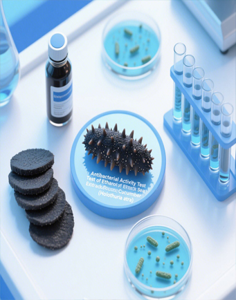
Antibiotic sensitivity testing is essential for determining bacterial susceptibility to antibiotics. In disc diffusion testing, several technical factors influence the diameter of the inhibition zone, including incubation temperature, which must be carefully controlled to ensure the validity of test results. This study aims to determine the mean, difference, and analyze the diameter of the inhibition zone of the antibiotics, namely Clindamycin, Levofloxacin, Tetracycline, and Trimethoprim against Staphylococcus aureus on Mueller-Hinton agar media with incubation temperatures of 33°C, 34°C, 35°C, 36°C and 37°C for 18 hours. This research is observational, with a cross-sectional design. The primary data is 100 data on the diameter of the antibiotic inhibition zone, obtained by measuring the diameter of the inhibition zone with different incubation temperatures. The selection of antibiotics is based on the mechanism of action of antibiotics inhibiting bacteria, namely the cell wall or cell membrane that surrounds the bacterial cell; the pieces of machinery that make the nucleic acids DNA and RNA, and the machinery that produces proteins (the ribosome and associated proteins) with a range of inhibition zones based on Internal Quality Control CLSI. The data and the repeated measure statistical test will be processed univariately to determine the significance of the difference in the diameter of the formed inhibition zone using the ANOVA test. The measurement of the inhibition zone diameter on the incubation temperature variation showed a significant difference with a p-value of 0.000 for Levofloxacin, Tetracycline, and Trimethoprim, while the p-value of Clindamycin is 0.010. For the other antibiotics, Levofloxacin, Tetracycline, and Trimethoprim antibiotics, the higher the incubation temperature, the average diameter of the inhibition zone is condensed, while for Clindamycin the higher the incubation temperature, the higher the average diameter of the inhibition zone is the same. Incubation temperature and volume affect the diameter of the antibiotic inhibition zone in the disc diffusion method for the antibiotic sensitivity test. It can be concluded that incubation temperature affects the diameter of the antibiotic inhibition zone in disc diffusion tests. It is recommended for future standardized and precise testing conditions to ensure accurate and reliable antibiotic sensitivity results.
CLSI. (2020). Performance Standards for Antimicrobial Disk Susceptibility Tests. 30th ed. CSLI Supplement M100.Wayne, PA: Clinical and Laboratory Standards Institute.
Coleman, D., Waddell, S. J., & Mitchison, D. A. (2011). Effects of low incubation temperatures on the bactericidal activity of anti-tuberculosis drugs. Journal of Antimicrobial Chemotherapy, 66(1), 146–150. https://doi.org/10.1093/jac/dkq414
Fanayoni, A., Gelgel, K. T. P., & Suarjana, I. G. K. (2019). Uji Sensitivitas Bakteri Staphylococcus sp. asal Babi Penderita Porcine Respiratory Disease Complex terhadap Doxycycline, Kanamycin, dan Clindamycin. Jurnal Indonesia Medicus Veterinus, 8(4), 439-445.
Goyat, R., Singh, J., Umar, A., Saharan, Y., Kumar, V., Algadi, H., Akbar, S., & Baskoutas, S. (2022). Modified low-temperature synthesis of graphene oxide nanosheets: Enhanced adsorption, antibacterial and antioxidant properties. Environmental Research, 215, 114245. https://doi.org/10.1016/j.envres.2022.114245
Hong, S. K., Choi, S. J., Shin, S., Lee, W., Pinto, N., Shin, N., ... & Chong, Y. (2015). Establishing Quality Control Ranges for Antimicrobial Susceptibility Testing of Escherichia coli, Pseudomonas aeruginosa , and Staphylococcus aureus: A Cornerstone to Develop Reference Strains for Korean Clinical Microbiology Laboratories. Annals of Laboratory Medicine, 35(6), 635–638. https://doi.org/10.3343/alm.2015.35.6.635
Hudzicki, J. (2009). Kirby-Bauer disk diffusion susceptibility test protocol. American Society for Microbiology, 15(1), 1–23.
Jenul, C., & Horswill, A. R. (2019). Regulation of Staphylococcus aureus virulence. Microbiology spectrum, 7(2), 1-21. https://doi.org/10.1128/microbiolspec.GPP3-0031-2018
Karimela, E. J., Ijong, F. G., & Dien, H. A. (2017). Characteristics of Staphylococcus aureus Isolated Smoked Fish Pinekuhe from Traditionally Processed from Sangihe District. Jurnal Pengolahan Hasil Perikanan Indonesia, 20(1), 188-198. https://doi.org/10.17844/jphpi.v20i1.16506
Khan, M. S., Yang, C., Pan, H., Yang, K., & Zhao, Y. (2022). The effect of high temperature aging on the corrosion resistance, mechanical property and antibacterial activity of Cu-2205 DSS. Colloids and Surfaces B: Biointerfaces, 211, 112309. https://doi.org/10.1016/j.colsurfb.2021.112309
Kuvarina, A. E., Roshka, Yu. A., Rogozhin, E. A., Nikitin, D. A., Kurakov, A. V., & Sadykova, V. S. (2022). Antimicrobial Properties and the Effect of Temperature on the Formation of Secondary Metabolites in Psychrophilic Micromycetes. Applied Biochemistry and Microbiology, 58(3), 243–250. https://doi.org/10.1134/S0003683822030085
Leboffe, M. J., & Pierce, B. E. (2019). Microbiology: Laboratory Theory and Application, Essentials. Morton Publishing Company.
Lenggu, C. K. L., Indriarini, D., & Amat, A. L. S. (2020). Uji Aktivitas Ekstrak Etanol Kulit Daging Buah Lontar (Borassus Flabellifer Linn) Terhadap Pertumbuhan Escherichia Coli Secarain Virto. Cendana Medical Journal, 8(2), 96-107. Retrieved from: https://ejurnal.undana.ac.id/index.php/CMJ/article/view/3353
Li, W., Zhang, Y., Ding, J., Zhang, S., Hu, T., Li, S., An, X., Ren, Y., Fu, Q., Jiang, X., & Li, X. (2022). Temperature-triggered fluorocopolymer aggregate coating switching from antibacterial to antifouling and superhydrophobic hemostasis. Colloids and Surfaces B: Biointerfaces, 215, 112496. https://doi.org/10.1016/j.colsurfb.2022.112496
Lusher, P., Denyer, S. P., & Hugo, W. B. (1984). A note on the effect of dilution and temperature on the bactericidal activity of potassium sorbate. Journal of Applied Bacteriology, 57(1), 179–181. https://doi.org/10.1111/j.1365-2672.1984.tb02372.x
Mietzner, T. A., Jawetz, E., Melnick, J. L., & Adelberg, E. A. (2019). Jawetz, Melnick & Adelberg’s Medical Microbiology (28th edition). McGraw Hill.
Murray, P. R., Rosenthal, K. S., & Pfaller, M. A. (2021). Medical microbiology (9th edition). Elsevier.
Nadjamuddin, M., Ismail, I., Luthfi M. C.F.M., Fitriana, F., Rusli, R., ... & Mubarak, F. (2023). Pengantar Bakteriologi (1st ed.). Eureka Media Aksara.
Notoatmodjo, S. (2005). Metodologi penelitian kesehatan. Jakarta: Rineka Cipta.
Novard, M. F. A., Suharti, N., & Rasyid, R. (2019). Gambaran Bakteri Penyebab Infeksi Pada Anak Berdasarkan Jenis Spesimen dan Pola Resistensinya di Laboratorium RSUP Dr. M. Djamil Padang Tahun 2014-2016. Jurnal Kesehatan Andalas, 8(2S), 26-32. https://doi.org/10.25077/jka.v8i2S.955
Parte, A. C., & Smith, J. T. (1994). Effects of temperature on the bactericidal activities of ciprofloxacin and levofloxacin against Staphylococcus aureus and Staphylococcus epidermidis. Microbios, 79(319), 87–95. Retrieved from: https://pubmed.ncbi.nlm.nih.gov/7968664/
Pelczar, M. J. (2008). Dasar-dasar mikrobiologi. Jakarta: Universitas Indonesia.
Perdana, R., & Setyawati, T. (2016). Uji in-vitro sensitivitas antibiotik terhadap bakteri Salmonella Typhi di Kota Palu. Medika Tadulako: Jurnal Ilmiah Kedokteran Fakultas Kedokteran dan Ilmu Kesehatan, 3(1), 11-22
Ronzetti, M., Baljinnyam, B., Jalal, I., Pal, U., & Simeonov, A. (2022). Application of biophysical methods for improved protein production and characterization: A case study on an high‐temperature requirement A‐family bacterial protease. Protein Science, 31(12), e4498. https://doi.org/10.1002/pro.4498
Siregar, M. T., Wulan, W. S., Setiawan, D., & Nuryati, A. (2018). Kendali Mutu. Buku Ajar Teknologi Laboratorium Medik (TLM). Jakarta: Kementerian Kesehatan Republik Indonesia.
Smith, P., Finnegan, W., Ngo, T., & Kronvall, G. (2018). Influence of incubation temperature and time on the precision of MIC and disc diffusion antimicrobial susceptibility test data. Aquaculture, 490, 19–24. https://doi.org/10.1016/j.aquaculture.2018.02.020
Songnaka, N., Nisoa, M., Atipairin, A., Wanganuttara, T., & Chinnawong, T. (2022). Enhanced Antibacterial Activity of Brevibacillus sp. SPR19 by Atmospheric and Room Temperature Plasma Mutagenesis (ARTP). Scientia Pharmaceutica, 90(2), 23. https://doi.org/10.3390/scipharm90020023
Sun, R., Vermeulen, A., & Devlieghere, F. (2021). Modeling the combined effect of temperature, pH, acetic and lactic acid concentrations on the growth/no growth interface of acid-tolerant Bacillus spores. International Journal of Food Microbiology, 360, 109419. https://doi.org/10.1016/j.ijfoodmicro.2021.109419
Toy, E. C. (2008). Case Files: Microbiology, 2nd Edition. McGraw-Hill Medical.
Willey, J. M., Sherwood, L., & Woolverton, C. J. (2017). Prescott’s microbiology (Tenth edition). McGraw-Hill.
Winarwi, W. (2006). Uji Viabilitas Bakteri dan Aktivitas Enzim Bakteri Proteolitik pada Media Carrier Bekatul (sebagai Acuan Bahan Ajar Pokok Bahasan Virus, Monera, Protista di SMA) [Skripsi]. Surakarta: Universitas Sebelas Maret.
Yoon, J. Y., Yeom, W., Kim, H., Beuchat, L. R., & Ryu, J.-H. (2022). Effects of temperature, pH and sodium chloride on antimicrobial activity of magnesium oxide nanoparticles against E. coli O157:H7. Journal of Applied Microbiology, 133(4), 2474–2483. https://doi.org/10.1111/jam.15719
Zhang, Y., Zhao, X., Wang, H., Fu, S., Lv, X., He, Q., Liu, R., Ji, F., & Xu, X. (2022). Effect of Temperature on the Adhesion and Bactericidal Activities of Ag+-Doped BiVO4 Ceramic Tiles. Inorganics, 10(5), 61. https://doi.org/10.3390/inorganics10050061
Copyright (c) 2024 JURNAL INFO KESEHATAN

This work is licensed under a Creative Commons Attribution-NonCommercial-ShareAlike 4.0 International License.
Copyright notice
Ownership of copyright
The copyright in this website and the material on this website (including without limitation the text, computer code, artwork, photographs, images, music, audio material, video material and audio-visual material on this website) is owned by JURNAL INFO KESEHATAN and its licensors.
Copyright license
JURNAL INFO KESEHATAN grants to you a worldwide non-exclusive royalty-free revocable license to:
- view this website and the material on this website on a computer or mobile device via a web browser;
- copy and store this website and the material on this website in your web browser cache memory; and
- print pages from this website for your use.
- All articles published by JURNAL INFO KESEHATAN are licensed under the Creative Commons Attribution 4.0 International License. This permits anyone to copy, redistribute, remix, transmit and adapt the work provided the original work and source is appropriately cited.
JURNAL INFO KESEHATAN does not grant you any other rights in relation to this website or the material on this website. In other words, all other rights are reserved.
For the avoidance of doubt, you must not adapt, edit, change, transform, publish, republish, distribute, redistribute, broadcast, rebroadcast or show or play in public this website or the material on this website (in any form or media) without appropriately and conspicuously citing the original work and source or JURNAL INFO KESEHATAN prior written permission.
Permissions
You may request permission to use the copyright materials on this website by writing to jurnalinfokesehatan@gmail.com.
Enforcement of copyright
JURNAL INFO KESEHATAN takes the protection of its copyright very seriously.
If JURNAL INFO KESEHATAN discovers that you have used its copyright materials in contravention of the license above, JURNAL INFO KESEHATAN may bring legal proceedings against you seeking monetary damages and an injunction to stop you using those materials. You could also be ordered to pay legal costs.
If you become aware of any use of JURNAL INFO KESEHATAN copyright materials that contravenes or may contravene the license above, please report this by email to jurnalinfokesehatan@gmail.com
Infringing material
If you become aware of any material on the website that you believe infringes your or any other person's copyright, please report this by email to jurnalinfokesehatan@gmail.com.